
Advancement in X-ray technology on its way for Dr Yusuf Dadoo
The team from Dr Yusuf Dadoo Hospital recently celebrated World Radiology Day and dreams of advancing their own x-ray technology soon.
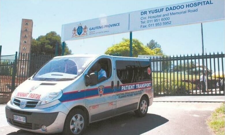
The radiology team from the Dr Yusuf Dadoo Hospital recently raised awareness about the benefits of radiographic imaging and therapy when they celebrated the lesser known World Radiology/ Radiographer Day.
A statement from the hospital’s Puseletso Mabidikame said the team also spoke about the value of radiology and radiologists’ contributions in the health system.
This year the team decided on the theme ‘From Roentgen to now’ and the hospitals CEO Dr Pheto was pleased about the concept of sharing the history of radiology and its evolution, which reaches beyond medical care.
“We see X-ray machines in many places across the country. Currently it is also being used in airports as part of crime prevention to make sure that drugs don’t move in the country,” he explained.
One of the hospital’s radiographers, Mechelle Christians, shared the history of X-rays and told attendees about physicist, Wilhelm Röntgen/Roentgen who was the first person to conduct an X-ray. The first ever X-ray was of his wife’s hand, a significant advancement that would ultimately benefit a variety of fields, most of all medicine by making the invisible visible.
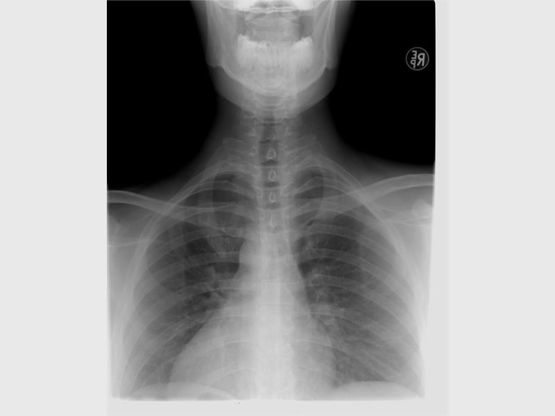
She also told them, “Our CEO’s desire is for us to go digital in the near future. With digital equipment you have the benefit of the machines having a reduced level of radiation for patients and staff, decrease film printing and switching over to electronic data.”
Another option is a Picture Archiving and Communications System (PACs). This is a computerised means of replacing the conventional X-ray film and connects to various work stations and department in the hospital. This means that doctors can view a patients X-rays from any station.
Mabidikame said in the statement that the Dr Yusuf Dadoo Hospital is currently in the beginning phase of this system.
“Then there is the new kid on the block, Artificial Intelligence, computer software that helps the Radiologist with diagnoses of the specific X-ray taken.”
She said that in the past the equipment was very bulky and complicated and developing X-rays in the dark was a process in its own. If the process was not done correctly it had to be repeated because it had no diagnostic value.
“X-ray equipment has become aesthetically more pleasing. Gone are the dark room days as computed radiography made its way into radiology.”
With the new technology came other advancements as well, like Fluoroscopy examinations which outlines soft tissue in the abdominal cavity, CT scans, Ultrasounds, Mammography, MRi’s and more.
Dr Johan Schutte also gave a lecture on radiology and the legal implications it could hold.